DEXA Scan as a Longevity Assessment Tool
DEXA scanning reveals body composition changes that weight and BMI cannot detect.
Weight and BMI cannot distinguish a pound of muscle from a pound of fat, and they cannot say where in the body that fat is sitting. That is the core limitation this piece starts from, and it is also the reason DEXA scanning has moved from a niche diagnostic tool into a serious longevity instrument. A scale gives a single number. BMI gives you a slightly more refined number, but it is still just height and weight. Neither one can separate lean tissue from fat mass, and neither can flag the handful of silent tissue changes that do the most to predict how someone ages.
The practical result is a kind of clinical blindness. Muscle loss, declining bone density, and rising visceral fat can each progress through early stages, the stages where intervention still works best, while the scale shows the same number and BMI stays in the same category. Vida Integrated Health frames this directly: these changes advance before any symptom appears. Someone can carry the same weight at 35 and at 55 while their body composition underneath has changed substantially, trading muscle for fat and losing bone density the entire time, with the scale reporting no news.
Consider two people, same height, same weight on the scale. One carries more muscle and stores fat subcutaneously. The other carries less muscle and stores a larger share of fat around the organs in the abdomen. Their metabolic profiles are not similar. Their disease risk is not similar. BMI treats them as identical, because it was never built to look past total mass. What the field needs instead is a tool that can separate fat from muscle, locate fat by region, and repeat the measurement reliably over months and years so a trend, not just a snapshot, becomes visible.
How a DEXA scan works
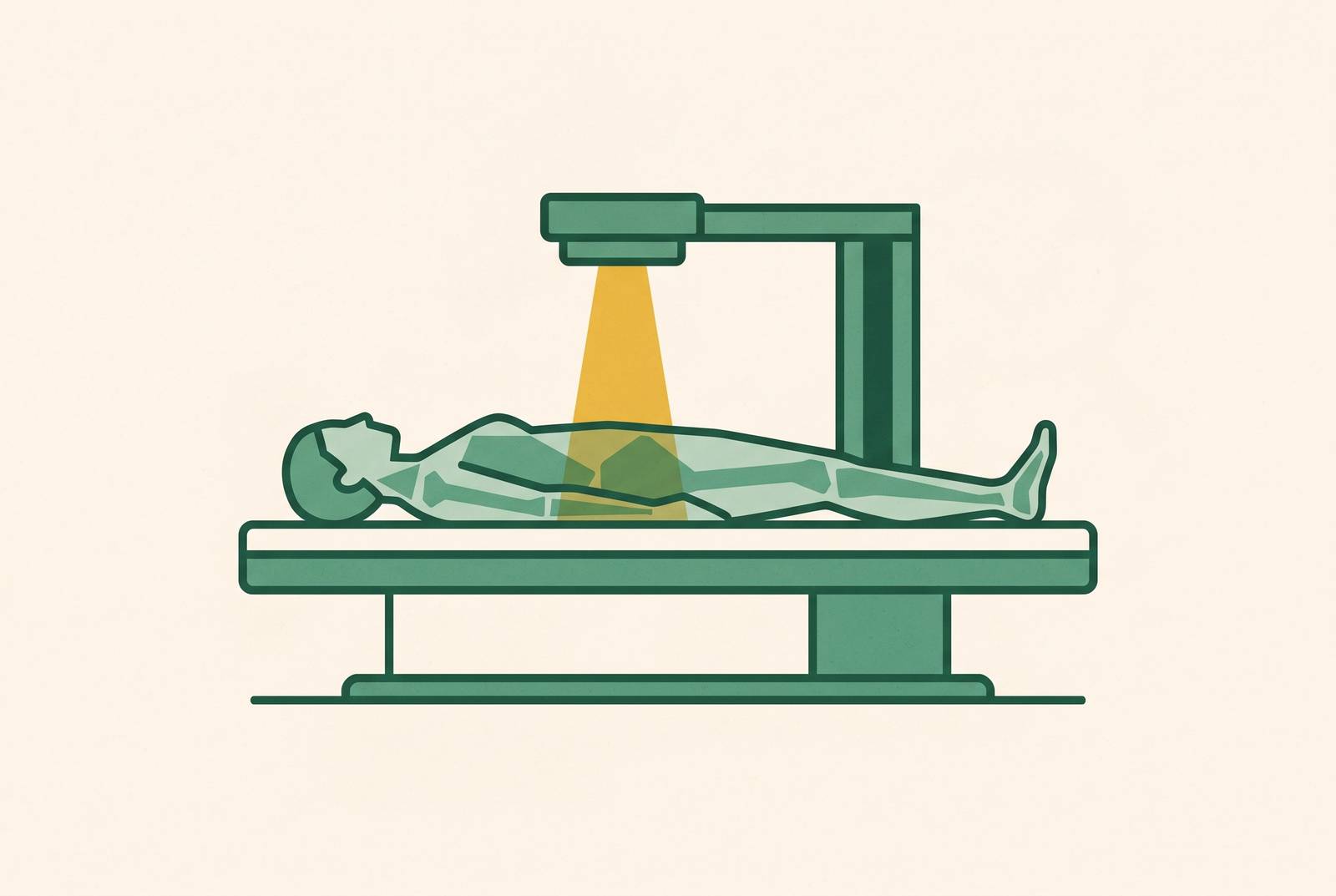
DEXA scanning solves that problem with a simple piece of physics. The machine passes two low-dose X-ray beams through the body, and because bone, fat, and lean tissue each absorb those two energies at different rates, the scanner's software can calculate how much of each tissue type is present. Vida Integrated Health describes this as a three-compartment model: bone mineral, fat mass, and lean soft tissue, measured separately rather than folded into one number the way height and weight are. That is a structurally different kind of output than anything a scale or a tape measure can produce.
The scan also reports by region, giving a single whole-body figure broken down by area. Dexaslim notes that the body gets segmented into arms, legs, and trunk, so the results show where lean mass is concentrated or missing and where fat has accumulated, rather than just how much of each exists in total. That regional detail turns out to matter enormously for the biomarkers covered later in this piece, since whole-body totals can look normal while a specific region is quietly declining.
Dexaslim says the radiation dose is low, under what you absorb from cosmic radiation on a standard cross-country flight, and that is part of why you can use repeat scanning as a routine screening tool rather than save it for rare diagnostic cases.
Two limits need stating before you go further, because they shape how you read every number in this piece. First, the lean mass figure DEXA produces includes water, skin, organs, and connective tissue, not skeletal muscle alone. Progevita, in guidance published August 21, 2026, flags this distinction directly: lean mass is not a synonym for muscle. Pair lean mass readings with a functional check like grip strength instead of treating them as a muscle score on their own.
Second, serial tracking requires consistency. Progevita's same guidance treats this as a practical precaution: comparing two scans only works if they come from the same scanner, the same protocol, and a known measurement error for that facility. If you switch machines between scans, the comparison can break entirely, turning what looks like a real change into noise from equipment differences.
DEXA began as an osteoporosis diagnostic, and it has since grown into full body composition analysis. Krakauer and Krakauer wrote in a peer-reviewed public health research journal that it holds status as a reference method for body composition assessment, because it is relatively non-invasive and can discriminate between tissue types. The rest of this piece builds on three specific numbers that method produces: visceral fat, limb muscle mass, and bone density.
Visceral adipose tissue: the fat DEXA finds that a mirror and a scale both miss
Not all fat behaves the same way inside the body, and that difference carries real metabolic weight. Fat stored just under the skin sits there fairly inert at normal levels. Fat stored around the liver, pancreas, and intestines, known as visceral adipose tissue, behaves more like an organ with its own agenda. Vida Integrated Health describes it as active fat, one that secretes inflammatory proteins and hormones that disrupt normal bodily function rather than simply taking up space.
The downstream effects run wide. DexaFit Scottsdale, in material published June 19, 2026, lists elevated systemic inflammation, insulin resistance, and increased cardiovascular and cognitive risk among the consequences tied to visceral fat specifically, distinguishing it from subcutaneous fat, which does not carry the same load. None of this appears on a scale or in a waist measurement taken alone. Someone's android-to-gynoid fat ratio can run high even when their total body fat percentage looks entirely reasonable, so they can look metabolically fine by every simple measure while still carrying a dangerous concentration of central fat. That gap is the clinical argument for measuring visceral fat directly instead of estimating it from weight or waist circumference.
Two honest limits apply here too. Progevita states that DEXA's visceral fat figure is an estimate, not a direct measurement. It correlates well with population data, but it works best read alongside waist circumference, blood pressure, fasting glucose, triglycerides, and ApoB rather than treated as a standalone verdict on cardiometabolic risk. There is no universal DEXA threshold that, by itself, diagnoses that risk. DEXA also cannot see inside organs. Liver fat and pancreatic fat require MRI or CT to measure directly, so an elevated visceral fat reading on a DEXA scan functions as a signal to look further, not as a complete metabolic audit on its own. Acknowledging that limit is part of what makes the case for using the scan well, rather than a reason to dismiss it.
The real value becomes visible over time. Progevita recommends treating visceral fat as one layer in a trend analysis, tracked on the same machine across repeat scans, rather than as a single number to react to. Even without a universal diagnostic cutoff, a visceral fat measurement that falls in response to a change in diet or training is a meaningful signal that something is working. Visceral fat predicts longevity risk on its own, apart from total body fat percentage, so testing this specific tissue metric gives you more actionable information than estimating body composition in general terms. Mito Health's membership model gives members access to DEXA scans at transparent, at-cost pricing, so they can capture measurements like this, the kind that weight and BMI cannot distinguish between. When platforms pair clinician-reviewed DEXA results with action plans built around visceral fat thresholds, you see a broader shift toward biomarker-driven preventive care that does not wait for symptoms to appear first.
Appendicular lean mass index: how DEXA detects sarcopenia before function declines
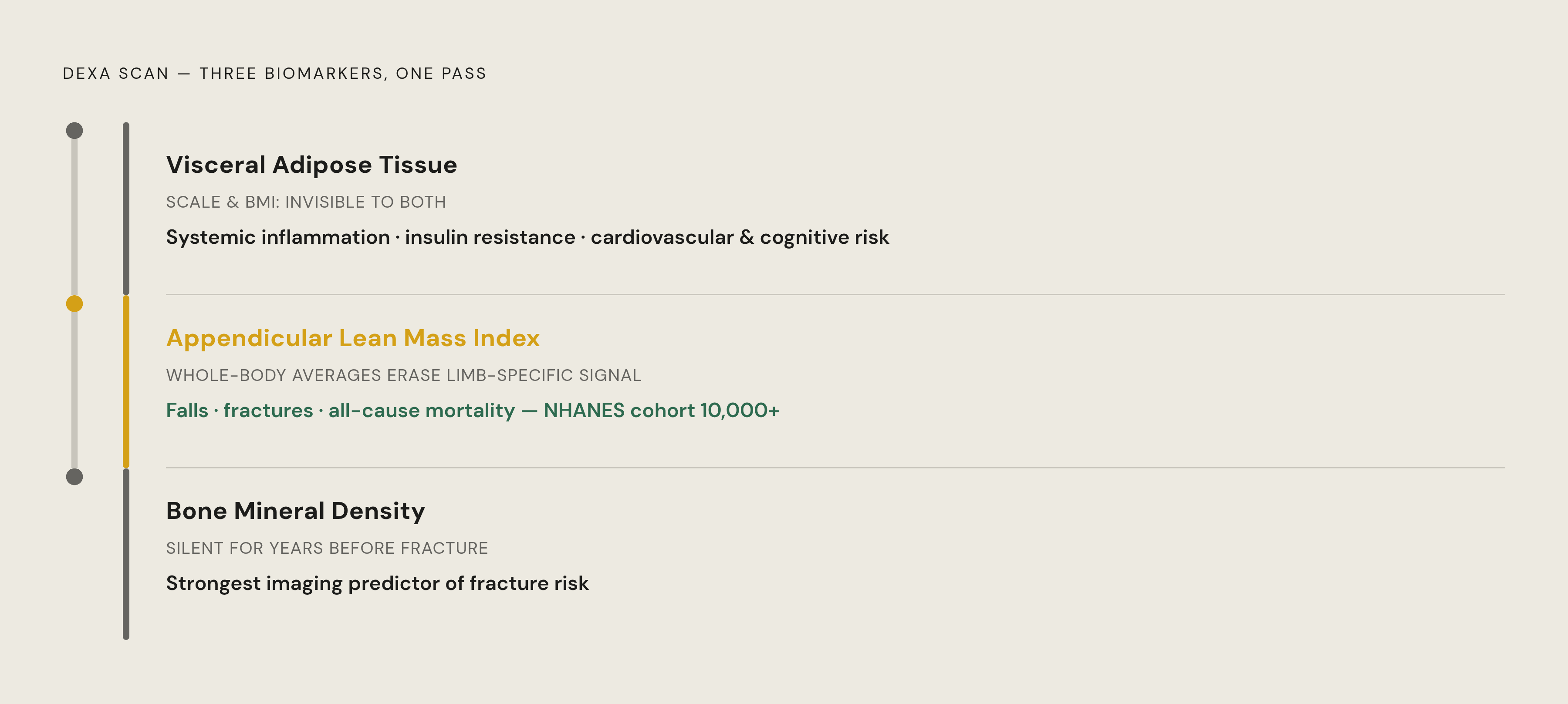
Muscle loss with age has a name, sarcopenia, and a dedicated clinical measurement, appendicular lean mass index, or ALMI. It sums the lean mass in the arms and legs and adjusts that figure for height, producing the specific number used in clinical sarcopenia diagnosis, a figure distinct from a rough proxy pulled from total body weight. DexaFit Scottsdale calls sarcopenia screening the most underappreciated insight a DEXA scan provides, and the stakes back that up: sarcopenia predicts falls, fractures, metabolic decline, and all-cause mortality. The CPH-MBD cohort protocol, from 2025, documents elevated risk of falls, fractures, and mortality tied to sarcopenia specifically in patients with stage 4 to 5 chronic kidney disease, one population where the stakes of missed muscle loss are especially high.
The research tying this regional measure to mortality is specific. Krakauer and Krakauer's 2021 analysis drew on an NHANES cohort of more than 10,000 participants, and it found that low limb fat-free mass, muscle mass in the arms and legs, was tied to elevated mortality risk. It did not meaningfully improve mortality prediction over basic anthropometrics like height and weight when taken alone. The regional number did. That is a striking result: it says the location of muscle loss carries information that a whole-body average erases.
DEXA results get their clinical usefulness from comparison. A 45-year-old's ALMI gets benchmarked against age- and sex-specific norms, DexaFit Scottsdale notes, so the question becomes whether that person's limb muscle mass is tracking ahead of, in line with, or behind their peers, rather than just whether it changed from last year. Sarcopenia itself is usually first suspected from reduced strength, with a DEXA number serving as confirmation rather than the initial signal. Progevita treats this distinction as practical rather than academic: ALMI confirms how much muscle is present, while grip strength and gait speed confirm how well that muscle functions. Both pieces belong in the full picture.
The reason early detection matters so much here is the shape of the decline itself. Muscle loss and reduced activity feed each other: less muscle makes movement harder, and reduced movement accelerates further muscle loss, building a cycle that gets harder to break the longer it runs. Catching the decline on a DEXA scan before it causes weakness or a fall gives someone a chance to intervene while reversal is still realistic, rather than after the cycle has taken hold. It is also worth noting that low bone density and low muscle mass tend to show up in the same people at the same time, a connection that sets up the next biomarker in this framework.
Tracking ALMI over repeat scans on the same machine turns it into a quantifiable longevity metric rather than a one-time data point. If you want to follow that trend, direct access to repeat scans, without needing a doctor's referral or an insurance company's sign-off each time, removes a real barrier to consistent tracking. That direct-access model is part of what Mito Health's testing approach is built around.
Bone mineral density: the fracture risk that builds invisibly over decades
Bone mineral density, or BMD, is the measurement DEXA was originally built to capture, and it is still the strongest predictor of bone strength and future fracture risk that imaging can give you. Osteoporosis is the condition that makes this measurement so valuable: it produces no symptoms for years, sometimes decades, until a fracture happens. That silent progression is exactly the pattern proactive screening exists to interrupt, the same pattern seen with visceral fat and muscle loss, just playing out in a different tissue.
DEXA reports bone density through two distinct scores, and Vida Integrated Health lays out what separates them. A T-score compares your bone density against a healthy young adult of the same sex, and doctors use it to diagnose osteoporosis and osteopenia. A Z-score compares your bone density against others of the same age and sex, so it can flag bone loss that is unusual even when you account for normal aging. Dexaslim confirms these two figures as the core output of a bone density scan, each doing a different job: one flags disease risk against an absolute standard, the other flags whether someone's bones are aging normally relative to their peers.
These three biomarkers, visceral fat, limb muscle mass, and bone density, rarely move in isolation from one another. Low bone density and low muscle mass tend to occur together, since the same hormonal and lifestyle factors that erode one often erode the other at the same time. A single DEXA scan captures all three in one pass, which is part of why it functions less like a one-time diagnostic and more like an ongoing instrument for watching the body's structural health change over years. Reading the three together, rather than any one in isolation, gives a clearer picture of healthspan risk than weight or BMI could ever produce on their own.